MRI SOMATOSENSATION DEVICE
The mouth is one of the most densely innervated parts of the body. Several sensory receptors transmit touch in the mouth to the brain.
Nevertheless, despite its sensory richness, somatosensation in the mouth remains poorly understood.
Using a novel pneumatic computer-controlled stimulation device the project aimed at the simulation-driven mapping of the intraoral tissues, particularly the teeth and the tongue.
This provides new insights into the sensory processing and neural representation of intraoral surfaces while also suggesting future possible practical applications in cognitive neuroscience and clinical neuropsychology

The apparatus that was used to deliver air puffs on teeth was composed of the following components: a portable stimulus computer, an air canister (Oxygen Cylinder Industrial Grade, BOC, Linde Group ©, London, United Kingdom), a pneumatic control module (Huang & Sereno, 2007), a Data Acquisition Box (DAQ USB6361 X series, National Instruments, Austin, Texas), 8 polytetrafluoroethylenes (PTFE) tubes (TYGON, Gobain performance plastics, Charny, France) and custom-designed gumshields
A94 were tasked with creating made-to-measure, MRI safe gumshield devices that could deliver said air puffs to the surface of the teeth.
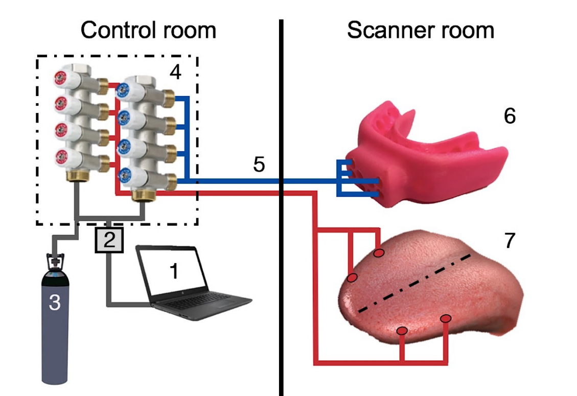
The figure above shows a detailed scheme of all the components of the pneumatic air puff stimulator used in the current study.
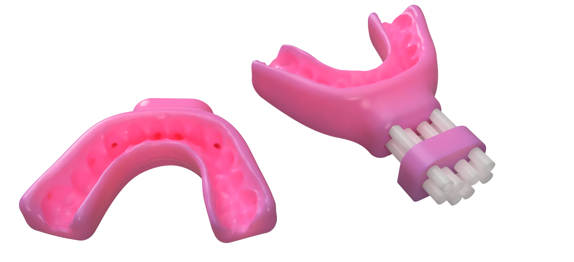
Controlled tactile stimulation of teeth with MRI compatible devices is not a trivial task. The non-noxious and computer-controlled nature of the stimulus was made possible by using a pneumatic air puff stimulator. In order to achieve somatotopic mapping of intraoral surfaces, we needed precise and highly localised stimulation (Medina & Coslett, 2010). The use of custom-designed 3D-printed mouthguards perfectly matched this need.
To allow stable fitting during stimulation, mouthguards were customised and individually produced for each participant based on their dental scans. Individual dental scans for both dental arches were acquired by using an intraoral scanner. This device allows non-invasive, accurate and direct capturing of the topography of human dentition

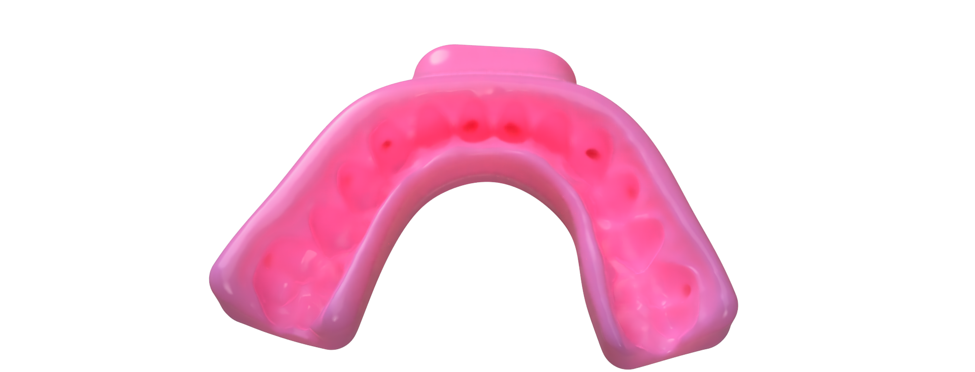
In this study, gumshields were used to safeguard the dissipation of the air puff stimulation across the dentition, thus delivering localised stimuli to teeth. Each gumshield was 3D printed by using a bespoke mouth-compatible material. For each participant, two gumshields were obtained, one for each dental arch. Both the mandibular and the maxillary mouthpieces had 8 internal channels. The starting point of each channel was located at the front side of the gumshield. In this location, each channel was connected and matched with one of the 8 PTFE tubes of the pneumatic air puff stimulator. The ending point of each intra-gumshield is channelled to the centre of the facial surface of a target tooth.